
Procedure for MRgFUS treatment
Step 1 – Pre-examination
After your personal gynecologist has diagnosed a fibroid using an ultrasound and determines that it is the most likely cause for the discomfort the patient is feeling, a MRT examination of the pelvis must be carried out at our FUS center or with a radiologist in the local area. After assessing the technical feasibility of the examination with the MRT images, you will be asked to attend a personal consultation and information meeting by the FUS specialist at our center or over the telephone if you have a long distance to travel.Step 2 – Defrayal of expenses and arranging an appointment
If you are suitable for treatment, you will receive an application from us for defrayal of expenses to be submitted to your health insurance company. Once we have received the treatment guarantee in writing, you will receive an appointment for MRgFUS treatment from our patient management. You will be sent written confirmation of your appointment along with all the information relating to your treatment.Step 3 – Day of treatment:
If the patients are receiving outpatient treatment, they will come to our FUS center in Dachau on the agreed date.You should eat a light breakfast at home, shave the hairs on your lower abdomen without using cream and then travel to the FUS center in Dachau.
After notifying the office of your arrival, you will have a detailed conversation where a demonstration will be given on the MRT images, the treatment procedure and the information sheet.
The patient is taken to the treatment room foyer where she can get changed in the dressing room (hospital gown). Intravenous access is established. This is necessary to be able to administer painkillers and sedatives. We also require a urinary bladder catheter to be able to monitor the filling level of the urinary bladder during treatment. Both of these items are provided by the doctor's assistant. There is another short conversation with the attending physician for any last questions.
The patient is then positioned as comfortably as possible in the MRT on a cushioned bed with in-built ultrasound device (pillows and blankets are provided). The patient lies with her head facing the outside and is able to see the medical workers through a pane of glass during treatment and is able to catch their attention by pressing an alarm ball or stop button. Ultrasound exposure can be interrupted at any time by pressing the stop button. Images are taken of the pelvic organs again to assess the current state of the fibroid. These images can be used to determine the best possible positioning for the patient which is easy to specify. The plan for uterine fibroid treatment will be prepared on the computer using the images, while the patient continues to lie still in the device. A mild sedative and painkiller ensures that the patient has very little awareness of the treatment and gives the impression that the time taken for said treatment is a lot shorter. All the points in the fibroid that are indicated are heated one after the other over a period of around 2-3 hours. The patient's pulse is controlled using a monitor and the temperature changes to the skin and in the body are displayed and controlled for every delivery of ultrasound.
After the last delivery of ultrasound, a final MRT is carried out using a contrast agent. This shows which areas of the fibroid are now no longer supplied with blood and the percentage of the uterine fibroid that has been treated. The area or the volume that is no longer supplied with blood is referred to as the "Non Perfused Volume" (NPV). Numerous international publications describe a NPV of over 60 percent as a good treatment result. Our comprehensive experience has shown that an NPV of over 80 percent is required to reduce the risk of relapse compared to a myomectomy. Please refer to our publications:
Magnetic Resonance Imaging-Guided Focused Ultrasound Treatment of Symptomatic Uterine Fibroids: Impact of Technology Advancement on Ablation.
Following treatment, the patient can lie back in the hospital bed, as they will be connected to another intravenous drip with glucose solution. The attending physician will discuss the treatment result with the patient and show them the images. The patient is then taken to the ward. She should stay there for another 1 to 2 hours. The staff nurse will also remove the urinary catheter and the venous access here. You are then able to leave the hospital accompanied by someone responsible (you are unable to drive due to the medication you have taken). It goes without saying that we provide our patients with the option to stay overnight at our hospital free of charge if they have a long journey home or if nobody is accompanying them.
The patient will be sent a treatment report and a CD with the MRT images in the next few days by post.
Step 4 – Check-up:
The treated fibroid will be partially or completely removed over a period of several months by your body's own immune system. The discomfort caused by the fibroid will decrease or gradually disappear completely. We recommend undergoing an MRT check-up 6 months after receiving treatment from your local radiologist or at our FUS center. You do not need to undergo any further examinations.Case example
Case example of a 47-year-old patient with multiple myoma nodes up to a diameter of 5 cm in size.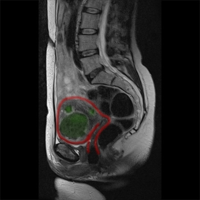
MRI images before treatment
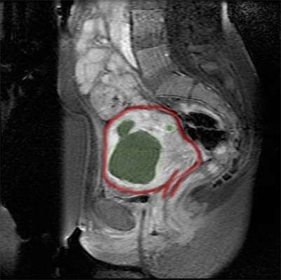
MRI images directly after the administration of contrast medium. The treated myomas are no longer perfused with blood, therefore they take up less contrast medium and remain black on the image.
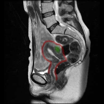
MRI images 11 months after MRgFUS treatment. The myomas are no longer visible or have shrunk markedly. The patient was completely free of symptoms 3 months after treatment.